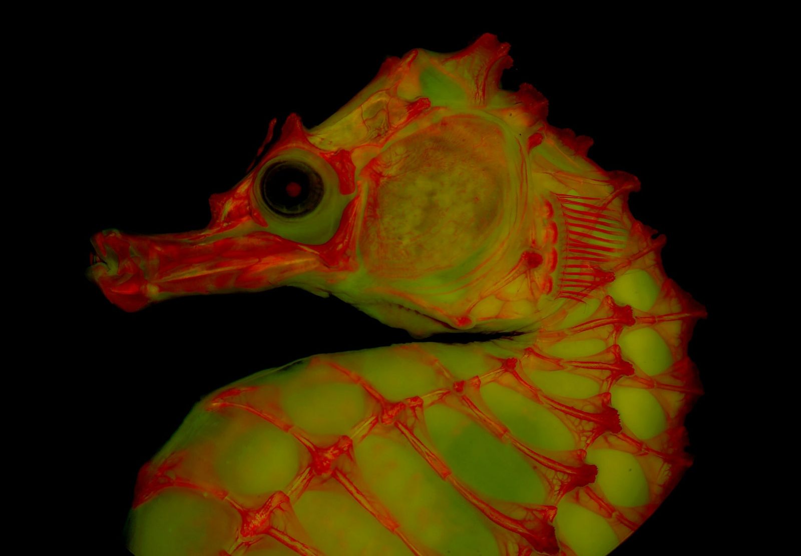
“There are so many different images that you can’t take,” says Leo Smith, a professor of ecology and evolutionary biology at the University of Kansas who helped develop the new technology. “If it’s a catfish, it’s going to settle on its stomach – that’s all it can do. If it’s a trout or something, it’s going to lie on its side because it collapses elsewhere.”
This is where gelatin comes in. Its gelatinous tissue can fix the skeletons in a position so that they can be photographed from different angles. After taking the photo, it can be washed again. This method, combined with the red dye and fluorescent light, enables recordings that were previously impossible.
The fluorescence microscope sees the red color
One evening in 2013, Smith – lead author for 2018 Published workWho describes this technique – a fishy skeleton colored under a fluorescence microscope on a whim.
“I held it underneath and thought, ‘What a holy straw, this is crazy, because the brilliance really showed every detail,’” Smith says.

Communicator. Reader. Hipster-friendly introvert. General zombie specialist. Tv trailblazer

